An Ultra-Rare Pediatric Cancer and the Intersectionality of Clinical Neglect
October 28, 2026
People

Evan Stoddard
ERI/PS2 Public Research Fellow
Evan Stoddard is a third year Ph.D. candidate in the Zeglis Laboratory at Belfer Research Building, where they study the development of radiotheranostics for a rare pediatric disease called desmoplastic small round cell tumors (DSRCT). Ava attended Appalachian State University and graduated in 2023 with their B.S. in molecular and cellular biology, before attending CUNY for graduate school. Ava is a 2026 ERI/PS2 Summer Fellow.
Many of us have had some interaction with the healthcare system in the U.S., and
therefore it’s probably safe to assume we have all in one way or another seen the
dangerous gaps and disparities that lie within it. Working in a pre-clinical academic
laboratory has situated me uniquely to see and address some of these gaps. The ERI/PS2
fellowship supported my dissertation research focused on developing diagnostic and
therapeutic tools for an ultra-rare pediatric cancer called desmoplastic small round cell
tumor (DSRCT). Roughly 20 people in the U.S. are diagnosed with DSRCT yearly, and its 5-
year survival rate is less than 15%. Furthermore, no standard-of-care has been established
for patients of this disease. This brings us to the first issue: due to its rarity, pharmaceutical
companies are not investigating diagnosis tools or treatments, because it isn’t profitable.
Even more so, the incidence rate of DSRCT among Black men is higher than other groups.
Like all things, the clinical gaps that patients of DSRCT face are intersectional, with
capitalism and medical bias at its center. Working within an academic laboratory allows
researchers to focus on more rare or neglected topics, which is at least partly why I am
passionate about this research.
Over the summer, I focused on finding biomarkers of DSRCT. Biomarkers are
proteins that are overexpressed – in my case, on the surface of cells – on the diseased cells
and are not expressed on healthy cells. Then, I utilize antibodies that target those proteins
to delivery radionuclides, such as zirconium-89 or leuticium-177, to the diseased cells.
This allows me to use Positron Emission Tomography (PET) to image the disease, or to treat
the cells with radioactivity. In the context of DSRCT, this would provide a non-invasive way
to diagnose and pinpoint the diseased cells and then would allow for the targeted
treatment of those same cells.
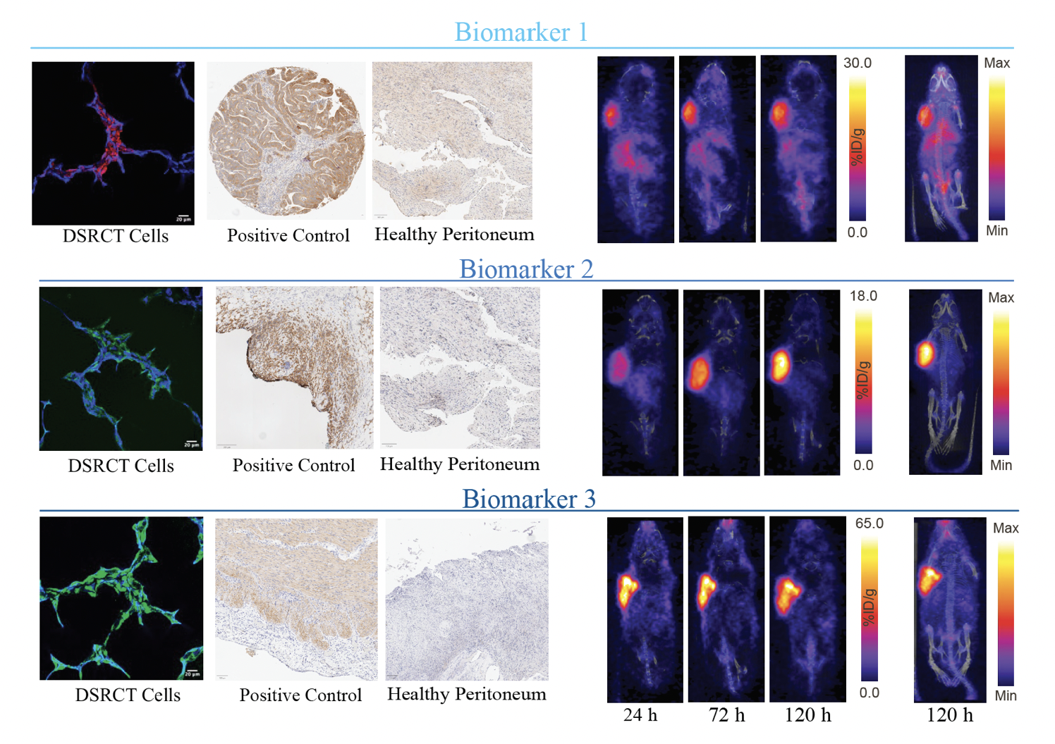
After doing literature research, I identified multiple potential biomarkers. I used
immunocytochemistry to image these biomarkers in DSRCT cells, and found high
expression of all three. DSRCT originates in the abdominal and pelvic peritoneal cavity, and
so I also stained healthy peritoneum tissue to confirm that these proteins weren’t
expressed. Using antibodies that target these three biomarkers, I was able to label them
with the radionuclide zirconium-89, which emits positrons and allows for PET imaging. Any
diagnostic or treatment tool that researchers are aiming to put into a human clinical trial
must first be tested in mice to ensure their safety and efficacy. So, utilizing mice that I
inoculated with DSRCT cells, I was able to PET image their tumors. Biomarker 1 was the
least effective to target, but both biomarker 2 and 3 provided highly contrasting PET
images.The first arm of this project was focused on PET imaging DSRCT for diagnosis and
patient stratification purposes, and the results of this project are currently under review for
publication! If you are interested in reading more, keep an eye out for that manuscript.
Looking forward, I am excited to continue my research on investigating safe and effective
treatments for DSRCT. No one should be neglected by the medical or scientific community
due to the rarity of their disease or due to their minoritized status. I am excited to have the
opportunity to continue my research with the support of programs like the ERI/PS2 Summer
Research Fellowship!